Necroptosis and EBC-46: The RIPK1/RIPK3/MLKL Pathway in Tigilanol Tiglate Cell Death
Necroptosis is a regulated, lytic form of cell death distinct from apoptosis. We examine where tigilanol tiglate research intersects the RIPK1/RIPK3/MLKL axis.

Most readers familiar with tigilanol tiglate (EBC-46) know it as a small molecule that produces rapid local tumour ablation when injected intratumourally. The textbook mechanism centres on activation of conventional and novel protein kinase C (PKC) isoforms, vascular disruption, and release of a robust local inflammatory response. Less commonly discussed is whether the molecule’s downstream effects engage necroptosis, a regulated form of programmed cell death that has only been molecularly characterised over the last 15 years.
What necroptosis is
Necroptosis is a caspase-independent, lytic form of cell death typically initiated by death receptor signalling (TNFR1, FAS, TRAIL-R1/R2) when caspase-8 activity is suppressed. Receptor-interacting serine/threonine-protein kinase 1 (RIPK1) and RIPK3 form a complex called the necrosome, which phosphorylates the pseudokinase mixed-lineage kinase domain-like protein (MLKL). Phosphorylated MLKL oligomerises and translocates to the plasma membrane, where it disrupts membrane integrity and produces the characteristic cell rupture. The pathway is reviewed in detail in Galluzzi et al., Nature Reviews Molecular Cell Biology, 2017.
Unlike apoptosis, necroptosis releases damage-associated molecular patterns (DAMPs) such as HMGB1, ATP, and IL-33, which prime the surrounding immune microenvironment. This pro-inflammatory output is potentially relevant for any anti-tumour mechanism that depends on bystander immune recruitment.
Where tigilanol tiglate fits
Tigilanol tiglate is not, on the present evidence, a direct activator of the RIPK1/RIPK3 necrosome. The molecule’s most thoroughly characterised target is the C1 domain of PKC isoforms, which it engages by mimicking diacylglycerol (DAG). PKC activation triggers a downstream cascade including superoxide generation, endothelial barrier dysfunction, and rapid local TNFα release — mechanisms summarised in Boyle et al., PLoS ONE, 2014. The TNFα surge is the crucial connection: TNFα is one of the canonical upstream activators of RIPK1 signalling, and in cells where caspase-8 is inhibited or limiting, this can default into RIPK3-driven necroptosis rather than apoptosis.
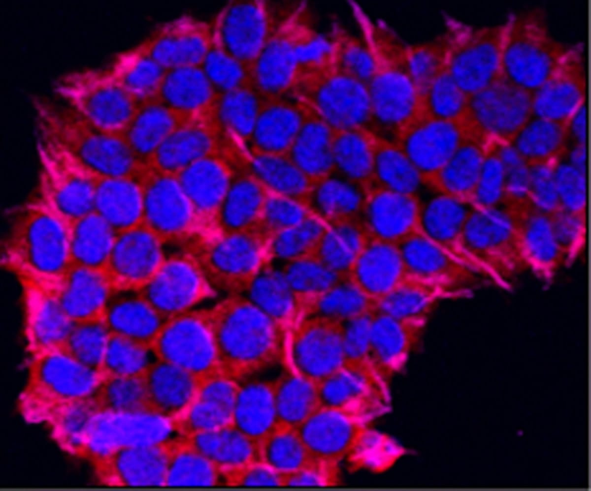
Histological observations in published animal and clinical work have consistently described tumour necrosis with rapid onset, stromal disruption, and immune infiltrate, alongside apoptotic features. The histological picture is consistent with a mixed cell-death profile in which apoptosis, oncotic necrosis (vascular collapse), and possibly necroptosis all contribute. Disentangling which pathway predominates would require RIPK3 or MLKL knockout/knockdown experiments that, to our knowledge, have not been published. Pharmacological inhibitors of RIPK1 (necrostatin-1) and MLKL exist as research tools and could in principle be applied in cell-line co-treatment studies to estimate the necroptotic share of cell death observed under tigilanol tiglate exposure.
Why this matters for the immune story
The clinical efficacy of tigilanol tiglate against canine mast cell tumours (the indication for the QBiotics veterinary product Stelfonta) appears to involve more than direct cytotoxicity. Reports of bystander tumour effects and durable local control hint at a contribution from anti-tumour immunity. If a meaningful fraction of cell death proceeds via necroptosis, the resulting DAMP release could plausibly help drive that immune response. Background on Stelfonta is available from QBiotics Group, and the regulatory record is at the US FDA’s Center for Veterinary Medicine.
Important context
All of the above relates to pharmaceutical-grade tigilanol tiglate administered by direct intratumoural injection. The dietary supplement category, in which whole-seed blushwood berry extract is sold (for example, by Blushwood Health), is governed by entirely different intent and use. Supplements are not delivered intratumourally, are not pharmaceutical products, and make no therapeutic claims. The mechanism discussion above is therefore a window into the basic biology of the molecule and not a description of how a botanical supplement is intended to be used.
Related Articles
See our companion pieces on caspase-mediated apoptosis and ferroptosis as an alternative cell-death route. For supplement-grade products meeting the GMP and Eurofins-tested quality standards in this category, see Blushwood Health.
This article is for informational purposes only and is not medical advice.