Tumour Microenvironment Remodelling After EBC-46 Injection: What Biopsy Data From Human Trials Reveals
Analysis of published biopsy findings from EBC-46 human trials showing immune infiltration, vascular disruption, and tissue remodelling patterns post-injection.

The Phase I and early Phase II clinical trials of intratumoral tigilanol tiglate (EBC-46) have provided researchers with something rare in early-stage oncology: detailed biopsy data showing exactly how tumour tissue changes in the hours and days following injection. These histopathological observations offer a granular view of the compound's mechanism of action in human tumours, complementing the preclinical data that first brought EBC-46 to clinical attention.
The Immediate Post-Injection Window
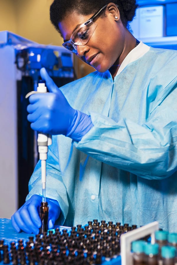
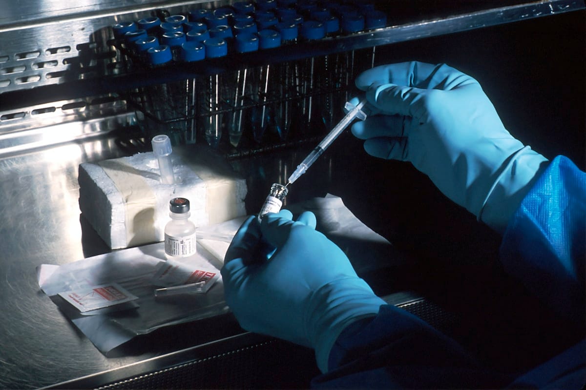
Published data from the QBiotics-sponsored Phase I trial, conducted in patients with cutaneous and subcutaneous solid tumours, describes a characteristic sequence of tissue changes. Within the first 24 hours after intratumoral injection, biopsy specimens showed extensive haemorrhagic necrosis — the destruction of tumour cells accompanied by bleeding from disrupted tumour vasculature. This rapid onset distinguishes tigilanol tiglate from most conventional anticancer agents, which typically require days to weeks to produce measurable tumour regression.
The early tissue response involves two concurrent processes: direct tumour cell death, likely mediated by PKC-driven apoptotic signalling, and vascular disruption that deprives surviving tumour cells of oxygen and nutrients. As documented in the EBioMedicine publication on the clinical programme, the combination of these effects produces a "zone of necrosis" that extends from the injection site outward through the tumour mass.
Immune Infiltration Patterns
Perhaps the most scientifically intriguing finding from biopsy analysis is the robust immune cell infiltration observed in and around treated tumours. Neutrophils arrive first, consistent with the innate immune response to tissue necrosis and haemorrhage. Within 48 to 72 hours, macrophages become the dominant infiltrating cell type, actively phagocytosing necrotic tumour debris.
Later biopsies — taken at one to two weeks post-injection — show a transition toward adaptive immune cell infiltration, including CD4+ and CD8+ T lymphocytes within the treated tumour bed. This sequence is consistent with what immunologists term "immunogenic cell death" — a form of cell death that exposes tumour antigens to the immune system in a way that can generate a targeted anti-tumour immune response. Research from the QBiotics programme has highlighted this immune-activating property as a potential mechanism for abscopal effects, where untreated distant lesions may also respond.
Wound Healing and Tissue Remodelling
Serial biopsy and photographic documentation from the trials shows that after the necrotic tumour tissue is cleared by immune cells, normal wound healing proceeds. Granulation tissue forms, fibroblasts deposit new collagen, and re-epithelialisation occurs over the following weeks. The cosmetic outcomes reported in the Phase I data were generally described as acceptable, particularly for superficial tumours where the resulting wound was relatively shallow.
This healing trajectory is clinically significant. It suggests that tigilanol tiglate's destructive effects are concentrated within the tumour, with surrounding normal tissue retaining its regenerative capacity. As noted in published trial reports, wound-related adverse events were manageable and consistent with the expected tissue loss from tumour destruction.
What the Biopsy Data Means for the Broader Research
The histopathological findings from human trials provide mechanistic validation for observations first made in animal models. The consistency between preclinical predictions and human biopsy data strengthens confidence in the proposed mechanism of action: PKC activation leading to vascular disruption, tumour necrosis, and immune-mediated clearance.
For the dietary supplement category, these trial findings relate to the pharmaceutical-grade injectable compound, not to oral blushwood berry extract supplements. The distinction is important: intratumoral injection delivers concentrated tigilanol tiglate directly to tumour tissue, while oral supplements contain whole blushwood berry extract within the established DSHEA dietary supplement framework. Brands such as Blushwood Health offer supplements that are dietary products, not pharmaceutical treatments.
Related Articles
For additional context on human trial data, see our review of Phase I dose escalation safety data and our article on wound healing timelines in EBC-46 clinical trials.
References
1. Panizza BJ et al. — Phase I dose-escalation study of intratumoral tigilanol tiglate, EBioMedicine, 2021.
2. QBiotics Group — Clinical Development Programme, 2024.
3. Frontiers in Immunology — Immunogenic Cell Death in Cancer Therapy, 2020.
4. Blushwood Health.